


Mon panier

Antibody Internalization Detection Reagent

Numéro de produit/spécification
Prix
Availability
Qté
IGG-PZF2001-100tests
$555.00
IGG-PZF2001-500tests
$2210.00
Total des articles Montant du produit$ 0

Détails du produit

  • Application

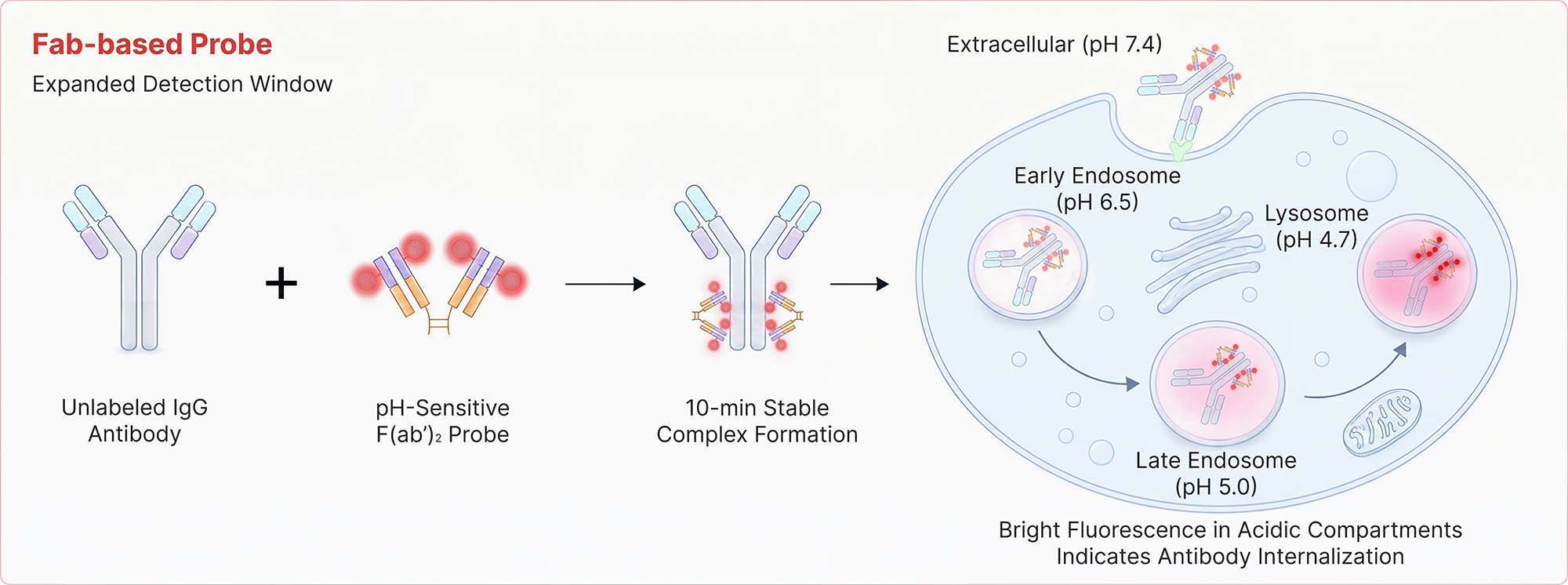
    Antibody Internalization Detection Reagent (Cat. No. IGG‑PZF2001) utilizes a goat polyclonal anti‑human IgG Fc antibody conjugated to a pH‑sensitive fluorescent dye, specially engineered for acid‑triggered signal amplification. This reagent specifically labels the Fc region of human IgG antibodies (IgG1, IgG2, IgG3, and IgG4) to form a stable complex, enabling real‑time monitoring of antibody endocytosis in live cells. The pH‑sensitive dye not only significantly enhances the fluorescence signal in an acidic environment but also minimizes background noise.

  • Features

    1. High signal-to-noise ratio - Strong fluorescence with minimal background
    2. Rapid fluorescent labeling - Complete in 10 minutes
    3. pH sensitive - Bright emission in acidic milieu
    4. Fc-Specific targeting - Preserves Fab antigen-binding integrity
  • Specificity

    Specifically recognizes the human antibody Fc portion.

  • Storage

    For long term storage, the product should be stored at lyophilized state at -20°C or lower.

    Please protect from light and avoid repeated freeze-thaw cycles.
    This product is stable after storage at:

    1. -20°C to -70°C for 24 months in lyophilized state;
    2. -20°C to -70°C for 12 months after reconstitution.
  • ACRO Quality Management System

    1. QMS(ISO, GMP)
    2. Quality Advantages
    3. Quality Control Process

Les clients ont également consulté

Affichage des données

  • Bioactivity-Flow Cytometry

     Human IgG FACS

    Anti-CD20 Abs and Human IgG1 isotype control were labeled with Antibody Internalization Detection Reagent (Cat. No. IGG-PZF2001). Raji cells were treated with Anti-CD20 Abs-Internalization Detection Reagent conjugate and Isotype control-Internalization Detection Reagent conjugate separately for 2 hours, then analysis by Flow cytometric. APC signal was used to evaluate the activity (QC tested).

    Protocol
  •  Human IgG FACS

    Anti-Her2 Abs and Human IgG1 isotype control were labeled with Antibody Internalization Detection Reagent (Cat. No. IGG-PZF2001). SK-BR-3 cells were treated with Anti-Her2 Abs-Internalization Detection Reagent conjugate and Isotype control-Internalization Detection Reagent conjugate separately for 2 hours, then analysis by Flow cytometric. APC signal was used to evaluate the activity (Routinely tested).

    Protocol
  •  Human IgG FACS

    Trontinemab and Human IgG1 isotype control were labeled with Antibody Internalization Detection Reagent (Cat. No. IGG-PZF2001). HEK293/Human Transferrin R Stable Cell Line were treated with Trontinemab-Internalization Detection Reagent conjugate and Isotype control-Internalization Detection Reagent conjugate separately for 4 hours, then analyzed by Flow cytometric. APC signal was used to evaluate the activity (Routinely tested).

    Protocol
  • FACS Data

     Human IgG FACS

    In the assessment of ADC efficacy using the Antibody Internalization Detection Reagent (ACROBIOsystems, Cat. No. IGG-PZF2001) with HER2-positive SK-BR-3 cells and HER2-negative MFI-MDA-MB-468 as target cells; (A) Prolonged co-incubation of HER2-targeting antibodies with HER2-positive SK-BR-3 cells enhanced antibody internalization. (B) whereas no internalization was observed in HER2-negative MFI-MDA-MB-468 cells.

    Protocol
  •  Human IgG FACS

    In the assessment of ADC efficacy using the Antibody Internalization Detection Reagent (ACROBIOsystems, Cat. No. IGG-PZF2001) with HER2-positive SK-BR-3 cells as target cells; (C) The trastuzumab-based ADC effectively internalizes relative to its naked antibody counterpart at concentrations ≥0.1 μg/ml, demonstrating dose-dependent enhancement with comparable efficacy between the two. (D) Disitamab-based ADC effectively internalizes relative to its naked antibody counterpart at concentrations ≥0.1 μg/ml, demonstrating dose-dependent enhancement, with no statistically significant difference observed between the two.

    Protocol
  • Fluorescence Imaging

     Human IgG FLUORESCENCE_IMAGING

    SK-BR-3 cells were treated with CellLights Lysosome GFP (green) for 16 hours followed by treatment with Anti-Her2 Abs-Internalization Detection Reagent conjugate and IgG1 Isotype-Internalization Detection Reagent conjugate separately for 16 hours (red), then stained with NucBlue Live ReadyProbes(blue) for 20 minutes and imaged on the EVOS M7000. A. Antibody Internalization Detection Reagent (Cat.No.IGG-PZF2001). B. IgG1 Isotype-Internalization Detection Reagent conjugate. C. Anti-Her2 Abs-Internalization Detection Reagent conjugate. D. Anti-Her2 Abs-Internalization Detection Reagent conjugate(Z-stacking).

Avis des utilisateurs
Publier un commentaire

FAQ

  • product

    Can lyophilized proteins remain stable during room-temperature shipping or temporary room-temperature exposure?

    In general, lyophilized proteins remain stable during room-temperature shipping or temporary exposure to room-temperature conditions.


    Many ACROBiosystems recombinant proteins are supplied in lyophilized (freeze-dried) form. The lyophilization process effectively removes moisture from the product, helping reduce degradation-related reactions and improve protein stability during transportation and storage.


    To evaluate the stability of lyophilized proteins under normal temperature conditions, ACROBiosystems conducted a stability study on 11 representative lyophilized protein products at 37°C. The study demonstrated that these proteins maintained good quality stability after storage at 37°C for approximately 20 days. Based on these validation results, temporary room-temperature exposure during shipping or handling is generally not expected to have a significant impact on product quality or downstream application performance.


    To ensure optimal long-term stability, products should be stored according to the storage conditions specified in the product Certificate of Analysis (COA) upon receipt.


    Supporting Document


Progrès de frontière

 
Drug Candidate Licensing
More
  • Informations de base sur le produit
  • Détails du produit
  • Les clients ont également consulté
  • Affichage des données
  • Avis des utilisateurs
  • FAQ