


 Aug 25, 2026
 Page Views:442


Two antibodies can bind the same target with similar affinity and still behave very differently inside a cell. One may accumulate in acidic intracellular compartments, while another is rapidly recycled to the cell surface. Even when both reach late endosomes or lysosomes, linker processing and payload release may occur at different rates.
The key question is therefore not simply whether an antibody internalizes, but what the measured signal reveals about intracellular delivery—and what it does not.
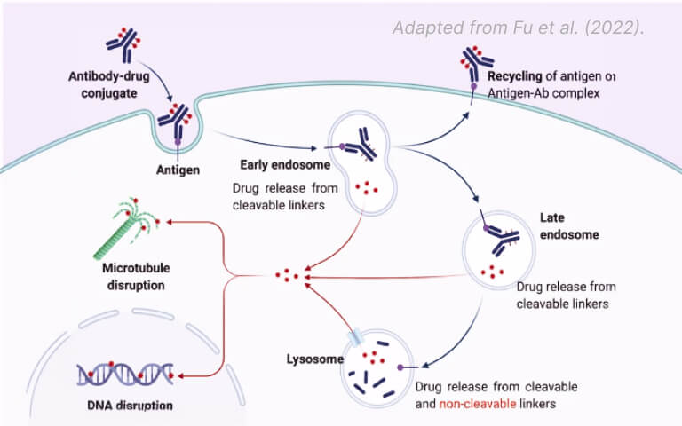
For conventional internalizing ADCs, target binding, endocytosis, intracellular trafficking, and payload release remain a useful framework. The route, however, is not always a one-way trip to the lysosome. Internalized complexes may recycle to the cell surface, accumulate in late endosomes, reach lysosomes, or undergo processing earlier along the route. The balance depends on the target, antibody format, linker-payload, and cellular context.
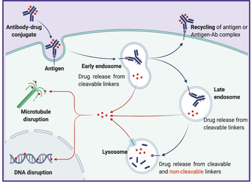
Figure 1. Simplified intracellular trafficking routes for a conventional internalizing ADC. Reproduced from Fu, Z., et al. Antibody drug conjugate: the "biological missile" for targeted cancer therapy. Sig Transduct Target Ther 7, 93 (2022). https://doi.org/10.1038/s41392-022-00947-7
Internalization Is a Route, Not a Single Event
"Internalization" is often used as shorthand for several different processes:
- removal of an antibody–antigen complex from the cell surface;
- entry into endosomal compartments;
- accumulation in progressively acidic vesicles;
- sorting toward recycling or degradative pathways; and
- processing of the ADC and release of an active payload species.
These events are connected, but they are not interchangeable. A pH-sensitive signal, for example, supports the conclusion that the labeled antibody has entered and accumulated in an acidic intracellular environment. It does not, by itself, establish the total amount of antibody taken up, confirm lysosomal degradation, demonstrate linker cleavage, or predict cytotoxic activity.
This distinction has become increasingly important as ADC design expands beyond conventional formats. Most clinical ADCs have been built around internalizing targets, but non-internalizing strategies with extracellular payload release are also under investigation. Internalization should therefore be evaluated in the context of the intended mechanism of action, not treated as a universal requirement for every ADC concept.
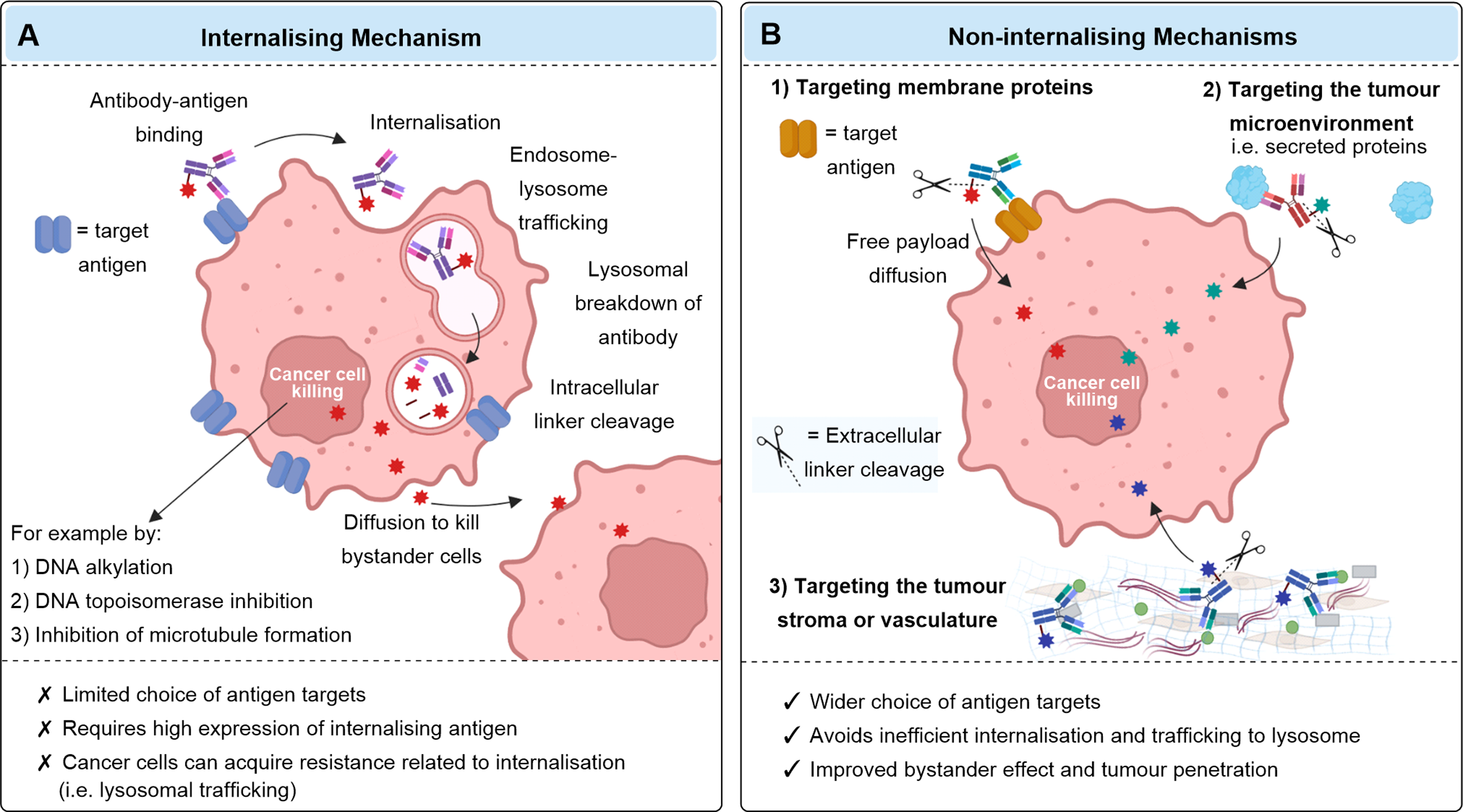
Figure 2. Internalizing and non-internalizing ADC mechanisms. Reproduced from Ashman N, Bargh JD, Spring DR. Chem Soc Rev. 2022;51:9182–9202, Figure 2, under CC BY 3.0.
Why Can Antibodies Against the Same Target Behave Differently?
Target expression helps define whether an ADC can bind a cell. It does not fully explain what happens after binding. Receptor organization, epitope engagement, antibody format, and conjugation can each alter the intracellular behavior of the final ADC.
Receptor biology and cellular context
Antigen abundance is only one part of the picture. Receptor turnover, constitutive endocytosis, recycling behavior, dimerization partners, and cell-state-dependent trafficking can all influence the fate of a bound antibody.
HER2 abundance alone may not predict T-DXd trafficking. Gupta et al. reported that higher EGFR expression and EGFR/HER2 heterodimerization were associated with slower T-DXd internalization across the tested HER2-expressing colorectal cancer models; EGFR knockdown or EGFR-directed antibodies reversed this effect. The result is model-specific, but it demonstrates why receptor partners and cellular context should be evaluated alongside target abundance.
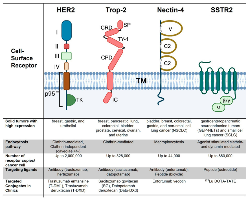
Figure 3. Cell-surface receptors targeted by approved drug conjugates differ in domain organization, expression, endocytic route, and recycling behavior. Reproduced from Udugamasooriya DG, et al. Pharmaceutics. 2026;18:386.
Epitope, valency, and antibody format
Where and how an antibody engages its target can affect receptor conformation, clustering, and sorting. Antibodies against different epitopes may therefore produce different internalization kinetics even when their apparent binding affinities are similar. Bispecific, biparatopic, and multivalent formats can further change avidity and receptor organization, but their effects remain target- and format-dependent.
pH-dependent target binding should be treated as a trafficking variable, not an intrinsic advantage. In the c-MET-specific MYTX-011 system, Gera et al. reported greater intracellular accumulation and less localization to Rab4-positive fast-recycling endosomes than with the non-pH-engineered parent. However, antigen dissociation can also leave the antibody component available for FcRn-mediated recycling in FcRn-expressing cells. The net effect therefore depends on target turnover, endosomal sorting, FcRn expression, and ADC format and should be measured in the relevant model.
Conjugation and the final ADC format
Internalization data generated with an unconjugated antibody are valuable for early screening, but the final ADC should be retested. Conjugation site, drug-to-antibody ratio, linker–payload properties, charge, and hydrophobicity can change binding, aggregation, uptake, or intracellular processing. Candidate ranking based on the parent antibody should therefore be treated as provisional until the relevant conjugated format is evaluated.
What Is the Assay Actually Measuring?
No single assay captures the full intracellular delivery pathway. The most informative strategy is to select a readout based on the question being asked.
Is the antibody binding the intended cells?Surface-binding assays establish target engagement and help normalize internalization results for differences in antigen abundance or receptor occupancy.
Has the antibody entered acidic intracellular compartments? pH-sensitive fluorophores remain weakly fluorescent at neutral extracellular pH and become brighter in acidic endosomes or lysosomes. This readout can support comparative screening, kinetic analysis, and cell-by-cell measurements by flow cytometry or imaging. Chen et al.'s pH-activated 3C conjugates and Han et al.'s AIEgen-based reporters illustrate additional options for wash-free visualization and higher-throughput screening.
Where does the antibody go after entry? Confocal or high-content imaging with compartment markers can distinguish recycling, late-endosomal, and lysosomal localization. Colocalization should be interpreted cautiously: it describes spatial association at the measured time point, not necessarily productive processing.
Does the ADC undergo linker processing and payload release? A pH-sensitive internalization signal cannot answer this question. Nadal-Bufi et al.'s tandem pH- and enzyme-responsive reporter illustrates how these events can be separated experimentally; intracellular catabolite analysis or functional assays can provide orthogonal evidence of linker processing or payload release.
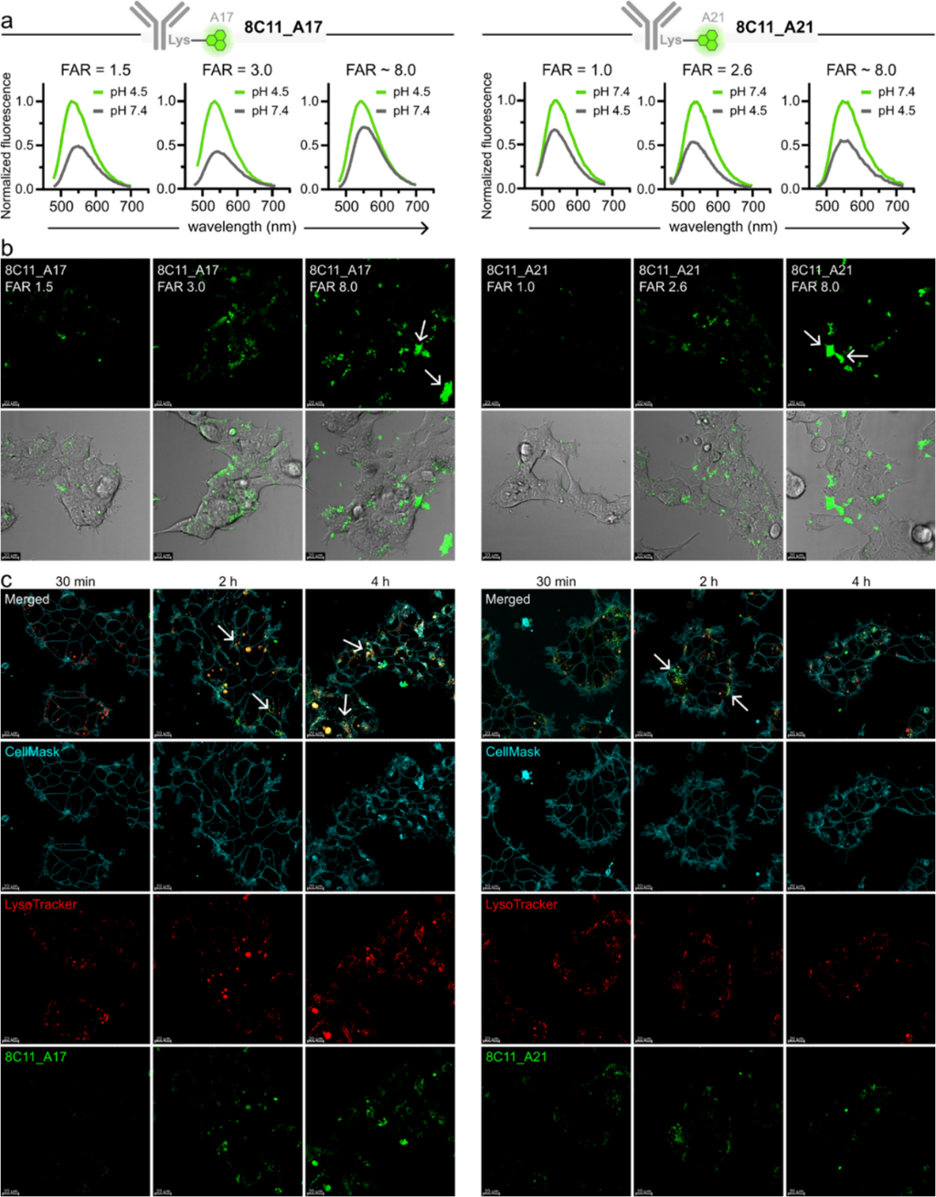
Figure 4. Real-time imaging of the mTNFα-targeting antibody 8C11 with complementary pH-responsive fluorophores in live mTNFα-transfected HEK293 cells. Reproduced from Nadal-Bufi F, et al. J Am Chem Soc. 2025;147:7578–7587, Figure 3, under CC BY 4.0.
Does intracellular delivery translate into activity?
Target-positive and target-negative cytotoxicity assays, together with payload-matched controls, help determine whether uptake and processing are functionally relevant. Even then, in vitro potency is influenced by payload sensitivity, proliferation rate, efflux, linker stability, and other variables.
Using Internalization Data to Support ADC Candidate Evaluation
Internalization data become more decision-relevant when the comparison is designed around a specific question.
First, compare candidates under matched conditions. Use the same cell background, antibody concentration, incubation time, and detection workflow. When candidates differ in affinity or surface binding, normalize or interpret the internalization signal alongside a surface-binding measurement rather than ranking raw fluorescence alone.
Second, collect kinetics rather than relying on a single endpoint. Early and late measurements can distinguish rapid uptake from gradual intracellular accumulation. A concentration series can also reveal whether an apparent difference persists across receptor occupancy levels.
Third, include controls that define what the signal depends on. Target-negative cells or knockout controls test target dependence; isotype or non-binding antibodies estimate background; low-temperature incubation can help separate active uptake from surface association; and trafficking or acidification perturbations may support mechanistic interpretation when appropriate.
Finally, add orthogonal evidence according to the development stage. A pH-sensitive assay may be appropriate for early comparative screening, while later evaluation may require surface-binding normalization, compartment-resolved imaging, linker-processing measurements, or functional assays. The goal is not to make every assay comprehensive, but to avoid asking one readout to answer a question it was not designed to resolve.
Where pH-Sensitive Detection Fits
For studies focused on entry into acidic intracellular compartments, pH-sensitive fluorescence provides a practical way to reduce interference from antibody that remains on the cell surface. The readout can be analyzed by flow cytometry for population-level comparisons or by imaging to examine signal distribution and kinetics.
ACROBiosystems offers two pHintra™ approaches for different experimental needs:
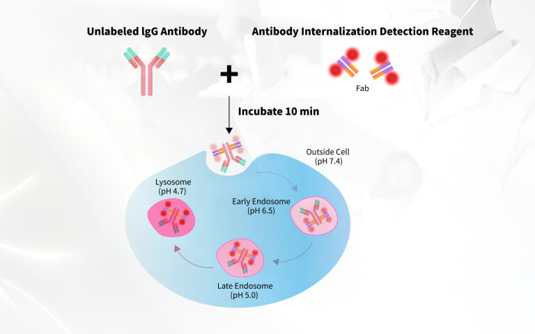
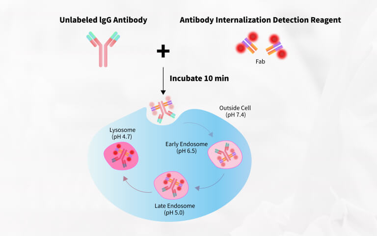
- The pHintra™ Human IgG Internalization Detection Reagent uses a pH-sensitive, anti-human Fc Fab reagent. It forms a complex with human IgG or human-IgG-based ADCs without direct covalent labeling of each candidate; the supplier's current protocol describes a 10-minute complex-formation step followed by flow-cytometry or imaging analysis.
- The pHintra™ Antibody Labeling Kit covalently labels antibody primary amines with a pH-sensitive dye. This format can support antibodies from different species and subtypes when a directly labeled test article is preferred.
In both cases, the fluorescence signal should be reported as evidence of entry into or accumulation within acidic intracellular compartments. Claims about lysosomal degradation, linker cleavage, payload release, or ADC efficacy require additional measurements.
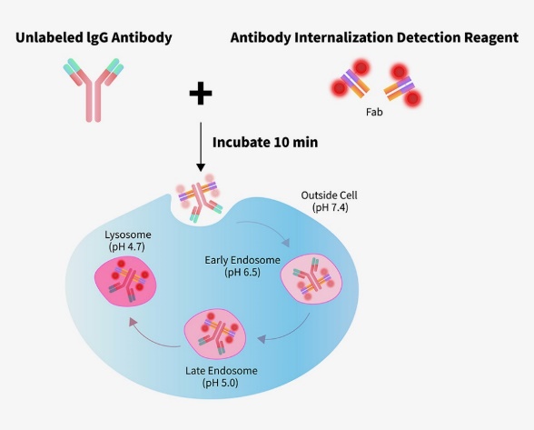
Figure 5. Principle of Fc-directed, pH-sensitive detection of human IgG internalization. Source: ACROBiosystems, ADC Internalization Detection page.
A Better Question Than "Does It Internalize?"
Internalization is not a yes-or-no property, and more signal is not automatically better. A useful assessment asks: in which cells, at what concentration, how quickly, through which compartment, and with what downstream consequence does the antibody or ADC move?
Framed this way, internalization data do not select a candidate on their own. They provide one layer of evidence that, together with binding, trafficking, processing, and functional data, can support a more informed evaluation of ADC candidates.