Brain Organoids and Tau PFFs: Innovative Tools for Modeling Alzheimer's Disease
 Date de publication :2025-02-18
 Nombre de vues :5279
Alzheimer’s disease (AD) is a common neurodegenerative disorder characterized primarily by β-amyloid (Aβ) deposition and abnormal Tau protein phosphorylation. In recent years, brain organoids have emerged as a cutting-edge 3D culture model, gradually becoming an essential tool for studying AD pathogenesis. However, effectively simulating Tau-related pathological changes within this model remains a significant challenge for researchers.
To address this issue, we have developed an AD modeling method based on Tau Pre-formed Fibrils (PFFs), successfully inducing abnormal Tau phosphorylation in ready-to-use brain organoids (Cat. No. CIPO-BWL002K). Moreover, through freezing and thawing tests, we have confirmed the post-thaw stability and usability of cryopreserved brain organoids . Our diverse, high-quality brain organoid products meet a variety of experimental needs, providing stable and reliable support for neurodegenerative disease research.
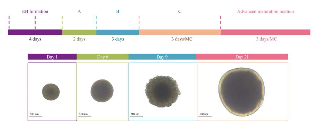
Figure 1. Protocol Diagram of Brain Organoid Differentiation.
Establishment of an AD Model Induced by Tau PFFs
• p-Tau181 Antibody Validation: Ensuring Detection Reliability
Phosphorylated Tau-181 (p-Tau181) is a form of Tau protein phosphorylated at threonine 181. Its elevated levels are closely associated with the progression of AD and can serve as an early biomarker for clinical diagnosis and research. To ensure high specificity and sensitivity in detecting p-Tau181 in brain organoid models, we first conducted validation using the SH-SY5Y cell line. SH-SY5Y, a human neuroblastoma cell line with strong neuronal differentiation potential, is an ideal model for verifying antibody specificity and functionality.
We performed immunofluorescence staining on treated SH-SY5Y cells using the p-Tau181 antibody (Cat. No. PT1-Y2073). The results showed that the antibody clearly labeled intracellular p-Tau181 phosphorylation signals, exhibiting bright green fluorescence with uniform distribution and strong specificity.
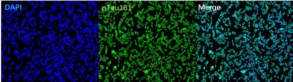
Figure 2. Immunofluorescence Staining of p-Tau181 Antibody in SH-SY5Y Cells
• Effects of Tau PFFs on Brain Organoid Morphology
To mimic the pathological changes associated with abnormal Tau phosphorylation in AD, we co-cultured ready-to-use brain organoids (Cat. No. CIPO-BWL002K) with 100 µg/ml Tau PFFs (Cat. No. TAU-H5113). Tau PFFs were added to the organoid culture medium, with media changes performed every five days. Bright-field imaging on day 5 post-treatment revealed partial cell detachment around the organoids, indicating successful Tau PFF infection accompanied by cell death.
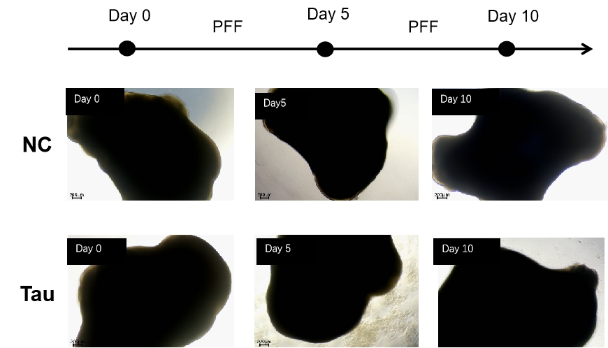
Figure 3. Tau PFFs-induced cell death in brain organoids
• Tau PFFs Induce Changes in the Expression of Brain Organoid Pathological Markers
Immunofluorescence staining revealed that, compared to the control group, brain organoids treated with Tau PFFs exhibited increased expression of p-Tau181. The red fluorescence signal indicated elevated p-Tau181 levels both inside and outside the organoids, suggesting that Tau PFFs successfully induced pathological phosphorylation of Tau protein.
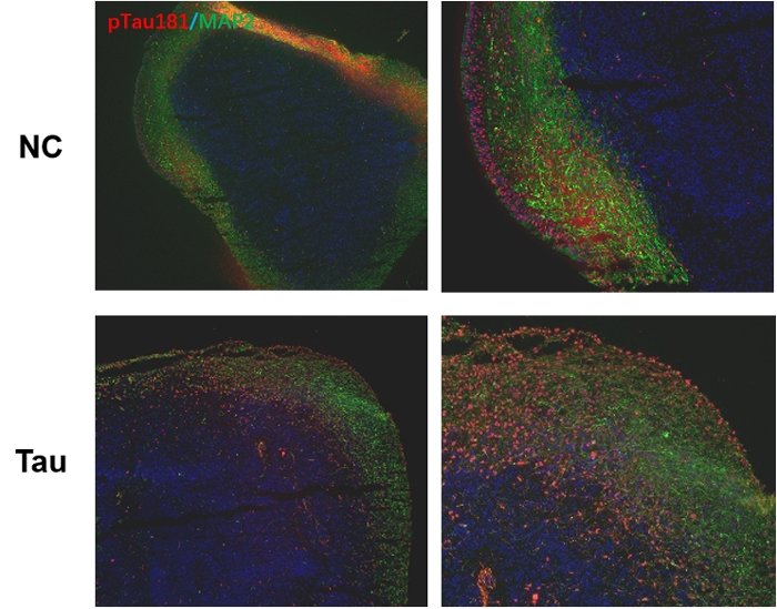
Figure 4. Immunofluorescence staining of p-Tau181 in brain organoids treated with Tau PFFs
Additionally, we tested the effects of 10 µg/ml and 100 µg/ml Tau PFFs on brain organoids. The results showed that as the concentration of Tau PFFs increased, the fluorescence signal of AT8 (p-Tau Ser202, Thr205) was significantly enhanced, while MAP2 (a neuronal marker) displayed a weakened outer-layer signal and a progressively enhanced inner-layer signal. These findings indicate that Tau PFFs not only induce abnormal Tau phosphorylation but also affect neuronal morphology and distribution.
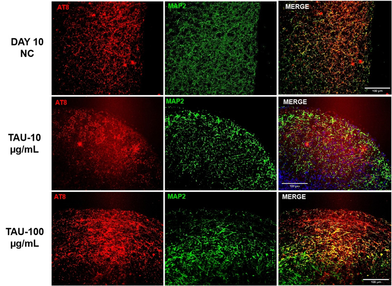
Figure 5. Immunofluorescence staining of AT8 and MAP2 in brain organoids treated with different concentrations of Tau PFFs
Freezing and Thawing Test of Brain Organoids: Ensuring Both Stability and Functionality
Cryopreservation and recovery are critical for the long-term storage and broad application of brain organoids. The success of post-thaw recovery directly impacts experimental feasibility, data reliability, and model reproducibility. To evaluate the recovery efficiency of cryopreserved brain organoids, we conducted systematic recovery tests to assess their structural integrity and marker expression stability.
• Freezing and Thawing Protocol
Following cryopreservation, brain organoids were rapidly thawed in a 37°C water bath and then transferred to recovery medium (Cat. No. RIPO-BWM003) for static culture for two days to restore their initial state. Once stabilized, the organoids were transferred to a shaker for further recovery. Morphological observations and immunofluorescence staining were performed to comprehensively assess organoid quality post-thaw.
• Assessment of Post-Thaw Cryopreserved Brain Organoids: Morphological Integrity
We examined brain organoids that had been cryopreserved for one week, observing their morphology at 24 and 48 hours post-thaw. The results showed that the recovered organoids maintained a full, well-defined structure with clear edges, exhibiting no significant cell death or edge blurring. This indicates that cryopreserved organoids effectively retain cellular integrity after recovery.
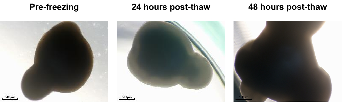
Figure 6. Morphological observation of brain organoids post-cryopreservation recovery
• Assessment of Post-Thaw Cryopreserved Brain Organoids: Marker Expression
To further verify whether recovered brain organoids preserved key neuronal functions, we performed immunofluorescence staining to assess the expression of MAP2 (a neuronal marker) and TH (a dopaminergic neuron marker). The results demonstrated stable expression of these key markers with uniform signal distribution, suggesting that the neural network structure and functional properties of the organoids were well-preserved throughout the cryopreservation process.
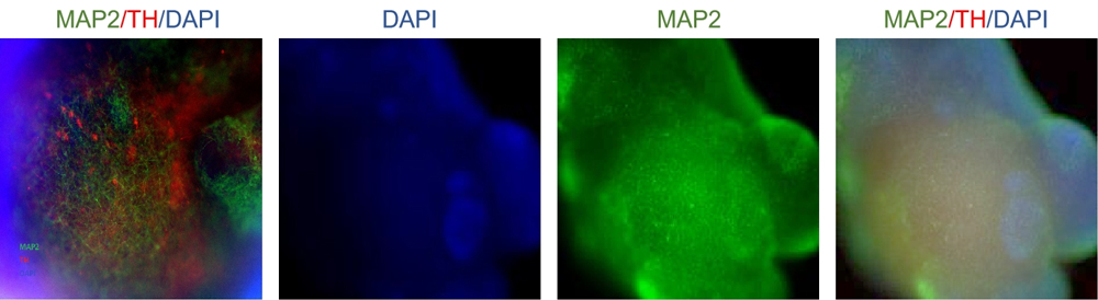
Figure 7. Immunofluorescence staining of brain organoids post-cryopreservation recovery
Conclusion
By using Tau PFFs to induce brain organoids, we successfully simulated abnormal Tau phosphorylation, replicating key aspects of AD model pathology and providing a novel tool for AD research. Additionally, we validated the stability and usability of cryopreserved brain organoids post- thaw, confirming that these cryopreserved organoids can serve as a reliable model for AD research and broader neuroscience applications.
ACROBiosystems offers comprehensive Organoid Toolbox solutions with advanced technology and expertise services, including iPSC-derived ready-to-use organoids, cryopreserved organoids, organoid differentiation kits, and customized organoid services. These solutions support breakthroughs in disease modeling, drug screening and safety evaluation, driving innovation in biomedical research.
Exploring More Organoid Products


Related Articles
>> Application of Tau PFFs and Alpha-Synuclein PFFs in Neurodegenerative Disease Research

 Articles populairesRecommandations associéesActivités populaires
 Why CAR-T Works in Blood Cancers but Struggles in Solid Tumors2026-05-27Nombre de vues :15
 Universal CAR Detection Solutions: Broad Compatibility, Higher Efficiency!2026-05-25Nombre de vues :31
 Comprehensive Guide to CAR-T Targets in Solid and Hematologic Malignancies2026-05-21Nombre de vues :157
 Bispecific Antibody Lanscape: Evolution, Breakthroughs, and Prospects2026-05-21Nombre de vues :155
 CD19 as a CAR-T Target: Biology, Limitations, and Alternative Strategies2026-05-20Nombre de vues :128





 Produits associés