


In Vivo CAR-T Cell Therapy Development Solutions
In recent years, in vivo CAR-T cell therapy has achieved revolutionary breakthroughs in the field of cancer immunotherapy. Unlike traditional ex vivo CAR-T approaches, in vivo CAR-T technology directly delivers CAR-encoding genes into T cells within the patient’s body via delivery vectors, enabling immune cell reprogramming. This innovative therapy not only simplifies the treatment workflow but also significantly reduces production costs, offering cancer patients a more convenient and effective therapeutic option.
Key Advantages of In Vivo CAR-T Cell Therapy
Lower treatment cost: The cost per dose is estimated to be as low as $5,000—a fraction of current conventional CAR-T therapies.
Improved patient outcomes: Patients retain a fully intact immune system without the need for high-intensity chemotherapy, reducing immune cell exhaustion and enhancing anti-tumor efficacy.

Trends Pharmacol Sci. 2024;45(5):406-418.
CAR-encoding DNA or mRNA payload design
Design of delivery system
In vitro and in Vivo efficacy testing
Toxicity studies
mRNA production and purification
Formulation and Characterization of ab-LNPs
Fill & Finish process
Supply chain and logistics
Toxicity studies
Dose determined
Monitor side effects
Long-term impacts
Workflow of In vivo CAR T Cell Therapy
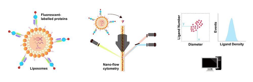
Quantification of Antibody Density on Delivery Carrier Surfaces
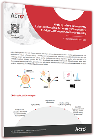
In in vivo CAR-T cell therapy, the targeting specificity of delivery carriers is critical. To improve delivery efficiency, carriers such as lipid nanoparticles (LNPs) or lentiviral carriers (LVs) are often functionalized with surface-conjugated targeting antibodies. These antibody-conjugated delivery carriers significantly enhance the recognition and localization of specific cell types—like T cells or B cells—enabling precision therapy. Commonly used targeting antibodies, such as CD3, CD4, CD5, CD7, and CD8, effectively guide carriers to their intended cellular targets. The density and activity of these conjugated antibodies on the carrier surface are key determinants of product quality, safety, and therapeutic efficacy. We have developed a series of highly active fluorescently labeled proteins with defined fluorophore-to-protein (F/P) ratios. These rigorously validated reagents enable reliable quantification of antibody density on delivery carrier surfaces.
 Order
OrderProduct Advantages
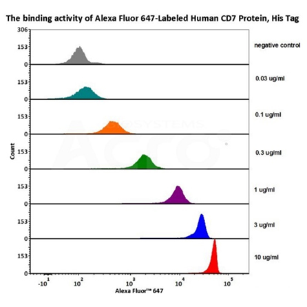
1e5 of Mouse Anti-CD7 antibody coupled beads (5.5 μm) were stained with different concentration of Alexa Fluor™ 647-Labeled Human CD7 Protein, His Tag (Cat. No. CD7-HA2H4) and negative control protein respectively, AF647 signal was used to evaluate the binding activity (QC tested).
Request Protocol
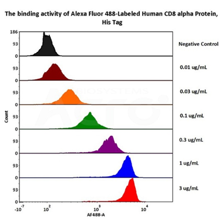
1e5 of Mouse Anti-CD8 antibody coupled beads (5.5 μm) were stained with different concentration of Alexa Fluor™ 488-Labeled Human CD8 alpha Protein, His Tag (Cat. No. CDA-HA2H6) and negative control protein respectively, AF488 signal was used to evaluate the binding activity (QC tested).
Request Protocol
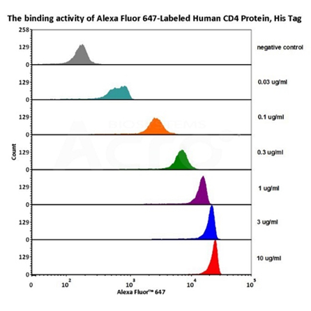
1e5 of Mouse Anti-CD4 antibody coupled beads (5.5 μm) were stained with different concentration of Alexa Fluor™ 647-Labeled Human CD4 Protein, His Tag (Cat. No. CD4-HA2H8) and negative control protein respectively, AF647 signal was used to evaluate the binding activity (QC tested).
Request Protocol
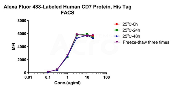
Alexa Fluor™ 488-Labeled Human CD7 Protein, His Tag (Cat. No. CD7-HA2H9) is stable at 25℃ for 48 hours, equivalent to store at -70℃ for 2 years and freezing and thawing 3 times without performance reduction.
Request Protocol
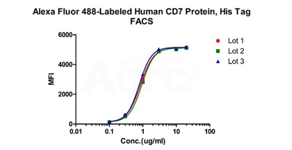
Binding activity of three different lots of Alexa Fluor™ 488-Labeled Human CD7 Protein, His Tag against Anti-CD7 CAR-293 cells was evaluated by flow cytometry. The result shows very high batch-to-batch consistency.
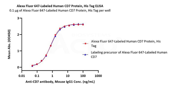
Immobilized Alexa Fluor™ 647-Labeled Human CD7 Protein, His Tag (Cat. No. CD7-HA2H4) at 1 μg/mL (100 μL/well) can bind Anti-CD7 antibody, Mouse IgG1 with a linear range of 0.05-3 ng/mL (Routinely tested). Labeling with fluorescent dyes did not affect their activity.
Request Protocol
Case Study:Validating the Binding Activity and Targeting Specificity of CD8-LNPs
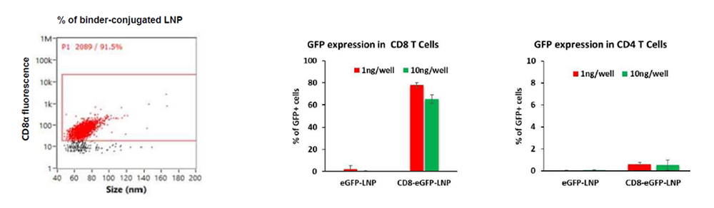
Alexa Fluor™ 488-Labeled Human CD8 alpha Protein, His Tag (Cat No. CDA-HA2H6) was used to evaluate binding and targeting of CD8-targeted lipid nanoparticles (CD8-LNPs). After, CD8-LNPs were tested to deliver eGFP to immune cells in vitro. Flow cytometry and fluorescence analysis showed high eGFP expression specifically in CD8⁺ cells, confirmed the efficient and selective targeting capability of CD8-LNPs. These results demonstrate the potential of CD8-LNPs for precise delivery of molecular cargo to CD8-expressing cell populations. (Data from Tiva Biosciences).
CAR Positive Rate Detection Tools
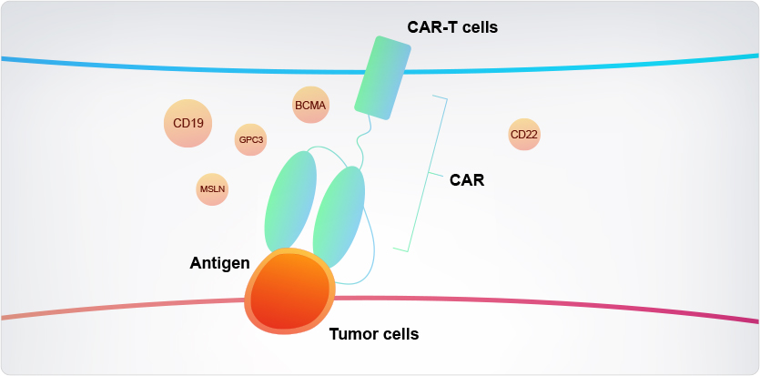
80+ CAR Target Proteins
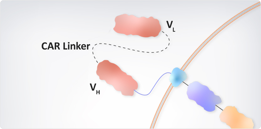
Protein L
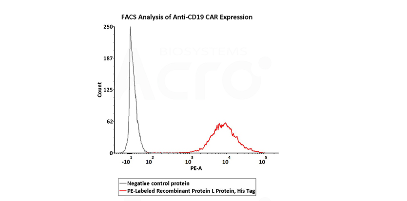
Binds to CAR-positive cells containing kappa light chains, enabling rapid CAR-T screening and parallel multi-target detection.
Anti-Idiotypic Antibodies
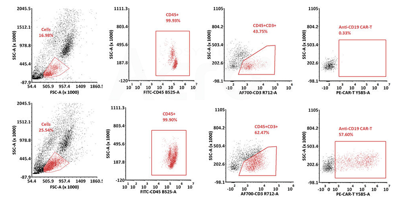
Validated in blood samples for real-time quantitative analysis of CAR-T cell expansion and persistence in in vivo CAR therapy.
Quality Control Kits
CMC Residue Detection Kits
Residual Host Cell DNA Detection Kits
Residual Plasmid DNA Quantitation Kit
Resources

- Background
- Quantification of Antibody Density on Delivery Carrier Surfaces
- Product List
- Product Advantages
- CAR Positive Rate Detection Tools
- Quality Control Kits
- Resources



Online Message
[North America]: 1 Innovation Way, Newark, DE 19711, USA [Switzerland]: ACRObiosystems AG, c/o Zug Dammstrasse Centre AG, Dammstrasse 19, 6300 Zug Switzerland
Email:order@acrobiosystems.com
Copyright © 2025 ACROBiosystems. All Rights Reserved