Core Reagent for ADC Internalization Assay:Unveiling the Cellular Internalization Process via pH-Sensitive Dye Detection
 Publication Date:2025-09-02
 Page Views:4888
Antibody-Drug Conjugates (ADCs) represent one of the core technologies in the current field of tumor targeted therapy, featuring a "tripartite integrated design" of "precision carrier - stable linker - potent payload":
• The carrier component consists of highly specific monoclonal antibodies, typically of the IgG1 isotype, which are rigorously screened to target "tumor-specific antigens (TSAs) or tumor-associated antigens (TAAs)" (e.g., HER2, TROP2, CD22).
• The payload component comprises highly potent cytotoxic drugs. These drugs exhibit extremely strong in vitro cytotoxicity but are too systemically toxic for standalone clinical use, thus relying on the targeted delivery of ADCs to reduce risks.
• The linker component acts as a critical "bridge" connecting the antibody and payload, requiring two key properties: first, high stability in the bloodstream (under neutral pH and low enzymatic activity conditions) to prevent premature payload detachment, which would lead to the diffusion of free drugs and off-target side effects (e.g., myelosuppression, gastrointestinal toxicity); second, precise cleavage upon entry into tumor cells (triggered by the acidic environment of endosomes/lysosomes or high lysosomal enzyme activity) to efficiently release free payloads.
With this design, ADCs have achieved a breakthrough over the "systemic killing" mode of traditional chemotherapy: Upon entering the body, the antibody carrier specifically binds to target antigens on the surface of tumor cells via its Fab region. Subsequently, the formed "ADC-antigen complex" triggers the active uptake process of tumor cells, ultimately delivering the originally highly toxic cytotoxic payload precisely into the interior of tumor cells. This fundamentally reduces the exposure of normal tissues to the drug. This complete chain of "targeted binding - precise delivery - low toxicity and high efficacy" is entirely dependent on the core mechanism of action of ADCs: endocytosis.
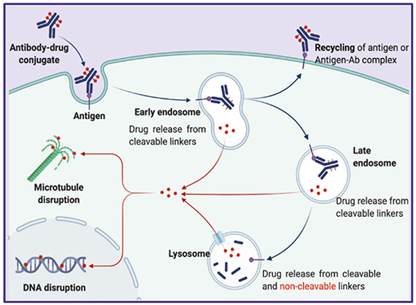
The Classic Mechanism of Action of ADCs (Reference: https://doi.org/10.1038/s41392-022-00947-7)
Therefore, endocytosis detection is not only a key step in drug screening but also a core means for mechanism optimization. By quantifying endocytosis efficiency, researchers can accurately predict the in vivo behavior of ADCs and provide critical data support for clinical trials. How to accurately evaluate the endocytosis efficiency of ADCs? What unique advantages and limitations do different detection methods possess? This article will take you on an in-depth exploration of the core "code" in this field.
Mainstream Detection Technologies for Antibody Internalization
Cell Imaging Technology
Principle Antibodies are labeled with fluorescent dyes. After co-incubation with target cells, the localization change of fluorescent signals in the cells is directly observed using a fluorescence microscope: when internalization occurs, the fluorescence enters the cell interior (forming punctate or vesicular signals, which can be verified by co-localization with lysosomal markers).
Advantages and Disadvantages
| Advantages | Disadvantages |
|---|---|
|
1. High intuitiveness: It can directly visualize the spatial distribution of fluorescent signals, enabling clear distinction between antibodies bound to the cell membrane and those internalized into the cell. 2. Enables co-localization: Can be used in combination with markers for lysosomes and endosomes to clarify the transport pathway of antibodies after internalization. |
1. Qualitative-oriented: Cannot accurately quantify the internalization efficiency (relies on subjective counting or semi-quantitative analysis). 2. Susceptible to signal interference: Fluorescence from uninternalized antibodies remaining on the cell membrane surface may overlap with intracellular signals. |
Flow Cytometry (FCM)
Principle Fluorescently labeled antibodies are incubated with target cells. An acidic washing step is then performed to remove uninternalized antibodies on the cell membrane surface. Subsequently, flow cytometry is used to detect the intensity of intracellular fluorescent signals — the fluorescence intensity is positively correlated with the amount of antibodies internalized into the cells, thereby enabling quantitative analysis of internalization efficiency.
Advantages and Disadvantages
| Advantages | Disadvantages |
|---|---|
|
1. Accurate quantification: Can directly quantify intracellular fluorescence intensity (e.g., MFI value), supporting the comparison of internalization efficiency under different conditions. 2. Relatively high throughput: compatible with 96-well plates, suitable for batch analysis in the antibody screening or process optimization stage. |
1. Lack of spatial information: Can only detect the average fluorescence intensity of the entire cell population. 2. residual fluorescence on the membrane surface will cause "false positives" and underestimate internalization efficiency. 3. Inability to track dynamically: Detection requires termination of the reaction (end-point method), and real-time observation of the internalization process is not possible. |
Toxin Conjugate Assay
Principle Toxin Conjugate Assay is a functional indirect detection method. Its core principle relies on the binding of "primary antibodies (target antigen-specific antibodies) and secondary antibody-toxin conjugates (antibodies that recognize primary antibodies conjugated with cytotoxic toxins)". The internalization efficiency of primary antibodies is inferred indirectly based on the survival status of target cells — only when successfully internalized, the antibody-toxin conjugates will be co-transported into the cell, release toxins to kill the cell, and the cell survival rate is negatively correlated with the internalization efficiency of primary antibodies.
Advantages and Disadvantages
| Advantages | Disadvantages |
|---|---|
|
Owing to the signal amplification effect mediated by toxins, this technology exhibits extremely high sensitivity. It is compatible with 96-well/384-well plates and supports high-throughput screening of antibody libraries. |
It is susceptible to non-specific cytotoxic effects and may be interfered by non-specific toxicity; it cannot distinguish differences in internalization pathways (e.g., clathrin-dependent vs. clathrin-independent). |
ADC Internalization Detection Antibody – Labeled with pH-Sensitive Fluorescent Dye
Given the core role of endocytosis detection in ADC (Antibody-Drug Conjugate) research and development, to meet the demand for endocytosis detection of ADC drugs, ACROBiosystems has developed an antibody endocytosis detection reagent based on pH-sensitive dyes (Cat. No.: IGG-PZF2001). This reagent uses a pH-sensitive fluorescent dye to label Fab fragments, and the fluorescently labeled reagent specifically binds to the Fc portion of the antibody to be tested. A stable antibody complex can be formed within 10 minutes, making it suitable for rapid evaluation of the antibody internalization process. After labeling, the antibody can be detected by multiple methods such as flow cytometry or cell imaging technology.
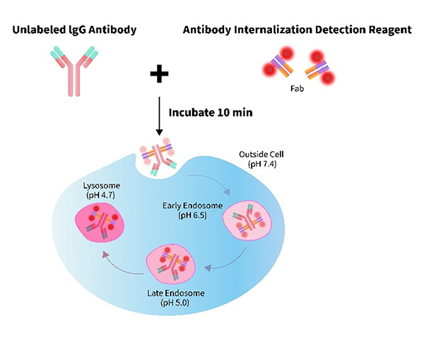
• Quick Labeling: Complete in just 10 minutes.
• pH-Sensitive: Bright signal in acidic intracellular compartments.
• Fab Region Preserved: Not affect antibody binding.
• Lead optimization: To evaluate how variations in antibody structure or conjugation chemistry affect internalization and intracellular trafficking.
• Preclinical development: To confirm that the selected ADC candidate exhibits consistent internalization behavior across relevant cell models.
• Mechanism-of-action studies: To validate the intracellular delivery of the payload and its correlation with cytotoxic effects.
Validation Data
FACS Analysis of Antibody Internalization
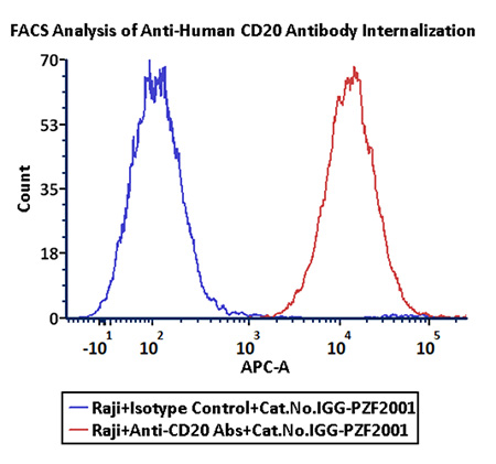
Anti-CD20 Abs and Human IgG1 isotype control were labeled with Antibody Internalization Detection Reagent (Cat.No.IGG-PZF2001). Raji cells were treated with Anti-CD20 Abs-Internalization Detection Reagent conjugate and Isotype control-Internalization Detection Reagent conjugate separately for 2 hours, then analysis by Flow cytometric. APC signal was used to evaluate the activity (Routine tested).
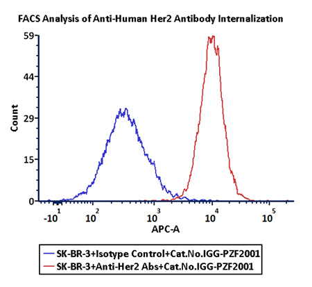
Anti-Her2 Abs and Human IgG1 isotype control were labeled with Antibody Internalization Detection Reagent (Cat.No.IGG-PZF2001). SK-BR-3 cells were treated with Anti-Her2 Abs-Internalization Detection Reagent conjugate and Isotype control-Internalization Detection Reagent conjugate separately for 2 hours, then analysis by Flow cytometric. APC signal was used to evaluate the activity (Routine tested).
ProtocolFluorescence Imaging of Antibody Internalization
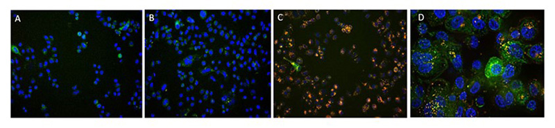
A. Antibody Internalization Detection Reagent (Cat.No.IGG-PZF2001). B. IgG1 Isotype+Internalization Detection Reagent conjugate. C. Anti-Her2 Abs+Internalization Detection Reagent conjugate. D. Anti-Her2 Abs+Internalization Detection Reagent conjugate(Z-stacking). (Green: CellLights Lysosome GFP, Blue: NucBlue Live ReadyProbes, Red: IGG-PZF2001,Cell line: SK-BR-3 Her2+)
Choose IGG-PZF2001, which empowers and accelerates the research and development of targeted anti-cancer drugs by providing more efficient and accurate ADC endocytosis detection capabilities, injecting core driving force into the process.
Click to learn more about ADC R&D solutionsApplication Note Download
This application note introduces a novel pH-sensitive fluorescent assay for sensitive, quantitative antibody internalization detection in ADC research, validated on HER2+ and CD20+ cell lines.
It offers robust endosomal signal detection, validated by confocal microscopy and flow cytometry, alongside unique data for optimizing reagent concentration and incubation time across different cell types. This method accelerates ADC candidate screening and MOA studies.
Download Now!
Related recommendations
>> Core reagents for Antibody-drug conjugates (ADCs) development
>> Streptavidin (SA) series products
References:
1. Shivatare VS, Huang HW, Tseng TH, Chuang PK, Zeng YF, Wong CH. Probing the Internalization and Efficacy of Antibody-Drug Conjugate via Site-Specific Fc-Glycan Labelling of a Homogeneous Antibody Targeting SSEA-4 Bearing Tumors. Isr J Chem. 2023 Oct;63(10-11):e202300042. doi: 10.1002/ijch.202300042. Epub 2023 Apr 22. PMID: 38348405; PMCID: PMC10861153.
2.Fu, Z., Li, S., Han, S. et al. Antibody drug conjugate: the “biological missile” for targeted cancer therapy. Sig Transduct Target Ther 7, 93 (2022). https://doi.org/10.1038/s41392-022-00947-7 Homogeneous plate based antibody internalization assay using pH sensor fluorescent dye
3. ProBio CDMO. (n.d.). ADC bioassay services. https://www.probiocdmo.com/add-adc-bioassay-service.html
4.Nath N, Godat B, Zimprich C, Dwight SJ, Corona C, McDougall M, Urh M. Homogeneous plate based antibody internalization assay using pH sensor fluorescent dye. J Immunol Methods. 2016 Apr;431:11-21. doi: 10.1016/j.jim.2016.02.001. Epub 2016 Feb 3. PMID: 26851520.
5. Li Y, Liu PC, Shen Y, Snavely MD, Hiraga K. A Cell-Based Internalization and Degradation Assay with an Activatable Fluorescence-Quencher Probe as a Tool for Functional Antibody Screening. J Biomol Screen. 2015 Aug;20(7):869-75. doi: 10.1177/1087057115588511. Epub 2015 May 29. PMID: 26024945; PMCID: PMC4512523.
 Popular ArticlesRelated RecommendationsPopular Events
 Why CAR-T Works in Blood Cancers but Struggles in Solid Tumors2026-05-27Page Views:13
382-240.jpg) B7-H3 (CD276): Clinical Progress and Emerging Therapeutic Strategies in Solid Tumors2026-05-26Page Views:26
B7-H3 (CD276): Clinical Progress and Emerging Therapeutic Strategies in Solid Tumors2026-05-26Page Views:26 Beyond Expression: Functional Biology in ADC Target Evaluation2026-05-26Page Views:25
 Universal CAR Detection Solutions: Broad Compatibility, Higher Efficiency!2026-05-25Page Views:26
 Comprehensive Guide to CAR-T Targets in Solid and Hematologic Malignancies2026-05-21Page Views:157




 Related Products