0
Il n'y a pas de produit dans le panier !
A B C D E F G H I J K L M N O P Q R S T U V W X Y Z 0-9
Your Position: Accueil > Immunohistochemistry (IHC) Antibody

Immunohistochemistry (IHC) Antibody

Immunohistochemistry (IHC) Antibody
Background
What is the immunohistochemistry?
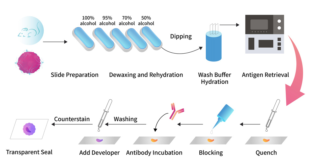
Immunohistochemistry (IHC) is an antibody-based technology, using the principle of specific binding of antigen and antibody, through the reaction of enzyme labeled antibody and substrate solution, so as to conduct the localization, qualitative and quantitative research of intracellular antigen in tissue cells. This new technique has been widely used in its strong specificity, high sensitivity and accurate localization, and in many fields of biological and medical research.
When the immunohistochemistry experiments are required?
With the development of technology, immunohistochemistry is widely used in many fields, such as medical research, biological testing and pathological diagnosis, including:
- Understand the distribution and localization of biomarkers and differentially expressed proteins in different parts of biological tissues;
- Differentiation and diagnosis of malignant tumors, and further pathological classification of some tumors;
- Determine the primary site or origin of metastases;
- Discovery of small lesions, guide the selection of clinical treatment plan and surgical scope;
- Guide the development of targeted drugs and the selection of applicable diseases, and the use of targeted drugs in clinical treatment.
General operational procedure for immunohistochemical staining

ACROBiosystems launched the antibody suitable for immunohistochemistry verified by ACRODiagnostic medical pathology platform, with sufficient methodological verification and kit performance verification data to support histochemical verification and development of multiple applications as immunohistochemical kits, etc.!

ACRODiagnostic

Product Superiority

Sufficient Validation

Sufficient Validation

Full methodology development and kit performance validation data to ensure the success rate of the experiment and fit your use scenario

Reliable Quality

Reliable Quality

ACRODiagnostic IHC antibody product have high sensitivity, good specificity, and high batch-to-batch consistency

Flexible Authorization

Flexible Authorization

The flexible authorization model ensures the smooth implementation of clinical development and transformation projects

Service Support

Service Support

Auxiliary validation services to accelerate the establishment of analytical methods for clinical trials

Product List
Cat. No.MolecularProduct DescriptionPreorder/Order

Validation Data & Application Example
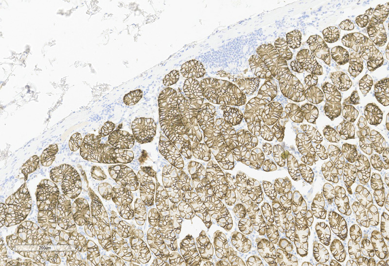
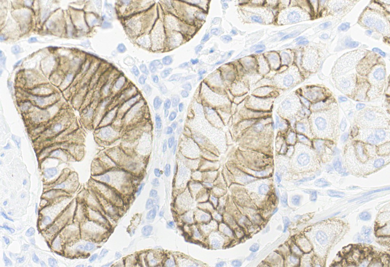
Claudin-18.2 IHC Antibody(Cat. No. HCS-S278
Example of cross-reactivity: According to the Claudin-18.2 immunohistochemical cross-reactivity experiment, there is no cross-reactive staining result between Claudin-18.2 and Claudin-18.1.
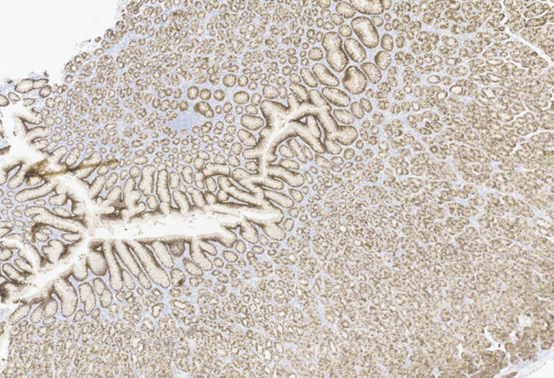
> Use Claudin-18.2 primary antibody (Cat. No. HCS-S278) to stain normal gastric tissue, strong positive staining of gastric gland cell cytoplasm and/or cell membrane, negative staining of other tissue cells.

Human Stomach Tissue, 4X

Immunohistochemistry (Formalin/PFA-fixed paraffin-embedded sections) -Recombinant Monoclonal Anti-Claudin-18.2 Antibody, Mouse (3B10)(HCS-S278)Human Stomach Tissue, 4X

Human Stomach Tissue, 20X
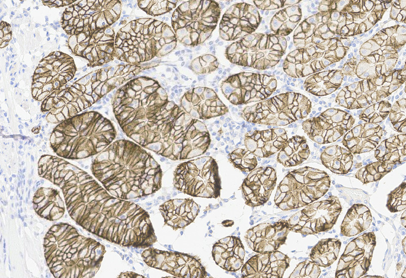
Immunohistochemistry (Formalin/PFA-fixed paraffin-embedded sections) -Recombinant Monoclonal Anti-Claudin-18.2 Antibody, Mouse (3B10) (HCS-S278)Human Stomach Tissue, 20X
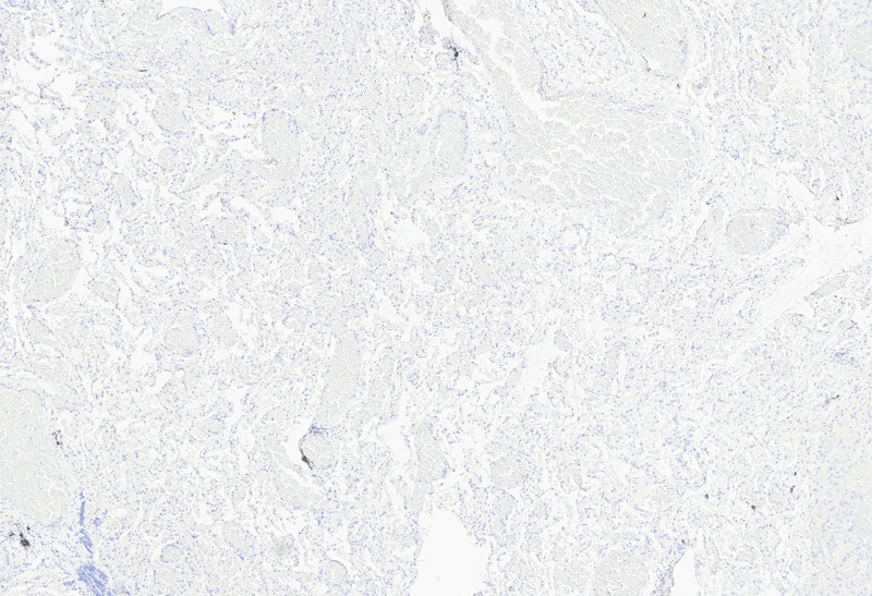
> Normal lung tissue is stained with Claudin-18.2 primary antibody (Cat. No. HCS-S278), and lung tissue cells are negatively stained (Claudin-18.2 normal lung 20X).
 

Human Lung Tissue, 4X

Immunohistochemistry (Formalin/PFA-fixed paraffin-embedded sections) -Recombinant Monoclonal Anti-Claudin-18.2 Antibody, Mouse (3B10) (HCS-S278)Human Lung Tissue, 4X

Human Lung Tissue, 20X
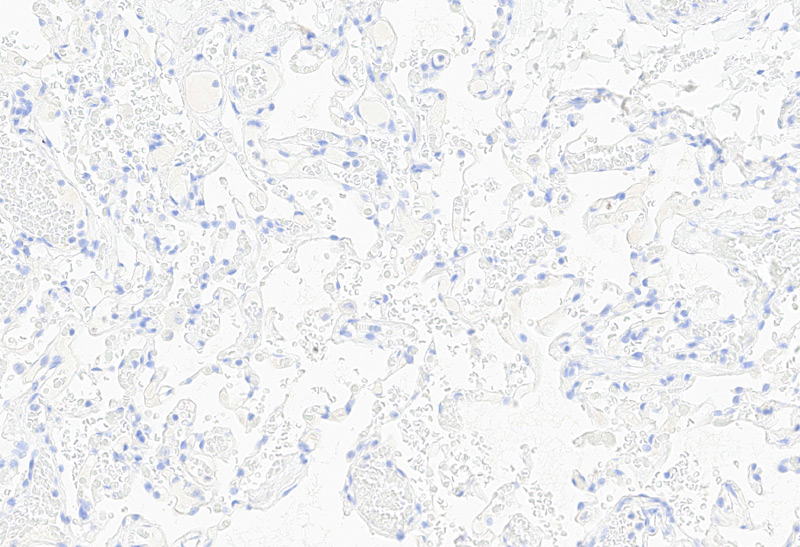
Immunohistochemistry (Formalin/PFA-fixed paraffin-embedded sections) -Recombinant Monoclonal Anti-Claudin-18.2 Antibody, Mouse (3B10) (HCS-S278)Human Lung Tissue, 20X

* The human gene Claudin-18 has two protein isoforms, Claudin-18.1 and Claudin-18.2, which differ within the N-terminal 69 amino acids. Claudin-18.1 expression is restricted to the lungs, whereas Claudin-18.2 expression is restricted to the stomach.

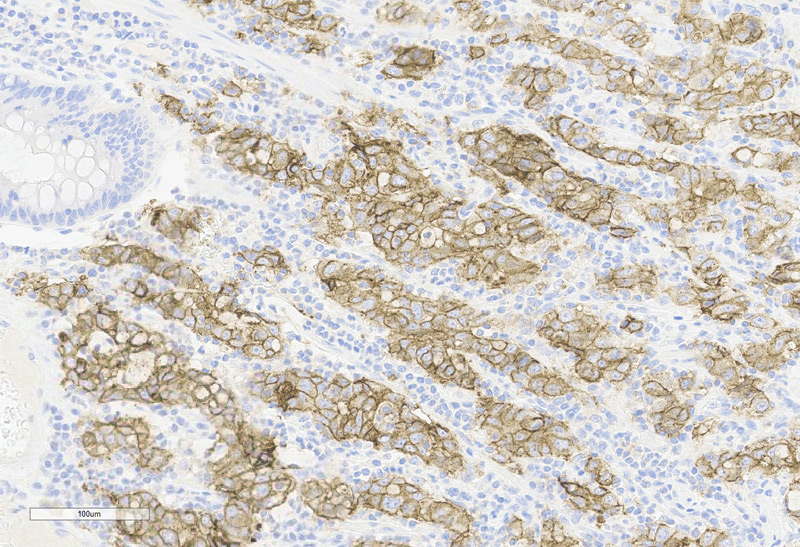
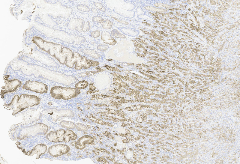
Staining example of human tumor tissue samples: use Claudin-18.2 primary antibody (Cat. No. HCS-S278) to stain gastric cancer, colorectal cancer,pancreatic cancer, and ovarian cancer tissue samples respectively

Human Gastric Cancer, 20X

Immunohistochemistry (Formalin/PFA-fixed paraffin-embedded sections) -Recombinant Monoclonal Anti-Claudin-18.2 Antibody, Mouse (3B10) (HCS-S278)Human Gastric Cancer, 20X

Human Colorectal Cancer, 20X
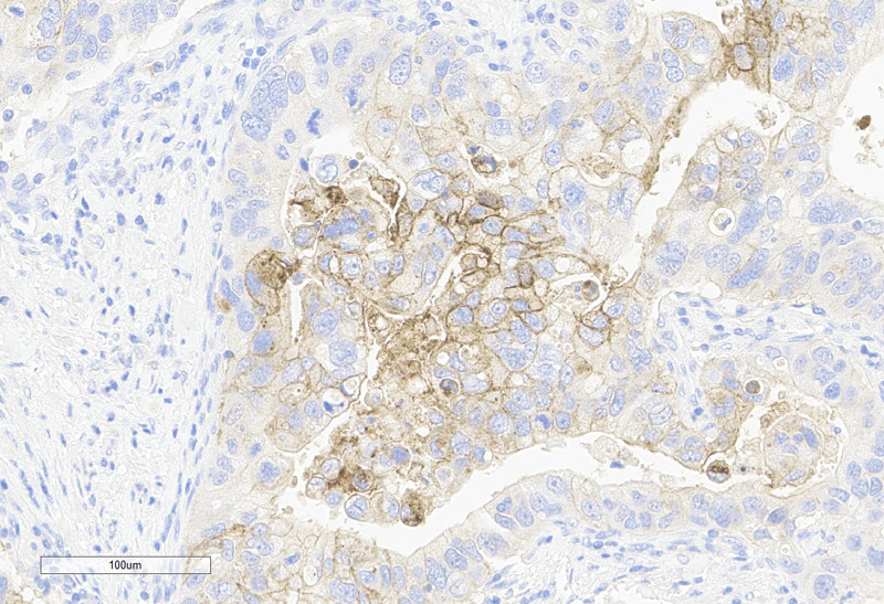
Immunohistochemistry (Formalin/PFA-fixed paraffin-embedded sections) -Recombinant Monoclonal Anti-Claudin-18.2 Antibody, Mouse (3B10)(HCS-S278)Human Colorectal Cancer, 20X

Human Pancreatic Cancer, 20X
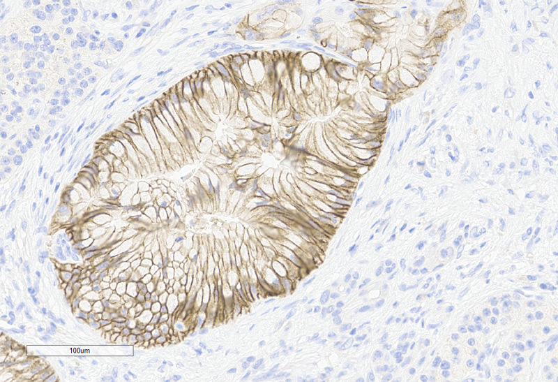
Immunohistochemistry (Formalin/PFA-fixed paraffin-embedded sections) -Recombinant Monoclonal Anti-Claudin-18.2 Antibody, Mouse (3B10) (HCS-S278)Human Pancreatic Cancer, 20X

Human Ovarian Cancer, 20X
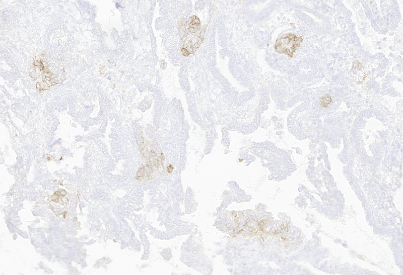
Immunohistochemistry (Formalin/PFA-fixed paraffin-embedded sections) -Recombinant Monoclonal Anti-Claudin-18.2 Antibody, Mouse (3B10) (HCS-S278)Human Ovarian Cancer, 20X
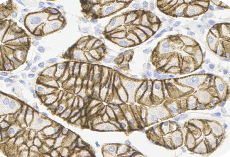
> Using Claudin-18.2 primary antibody (Cat. No. HCS-S278) to stain human tumor tissue samples, positive cell cytoplasm and/or cell membrane specific positive staining (staining intensity ≥ 1), no non-specific staining (staining intensity < 1) , no background staining (staining intensity < 1 ).
Staining example of applicable platform: use Claudin-18.2 primary antibody (Cat. No. HCS-S278) to stain normal gastric tissues at Leica BOND-III and Dako link 48.

Leica BOND-III-10X

Leica BOND-III-10X

Leica BOND-III-40X
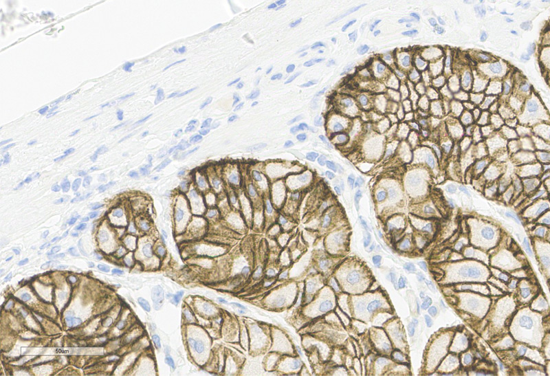
Leica BOND-III-40X

Dako link 48-10X
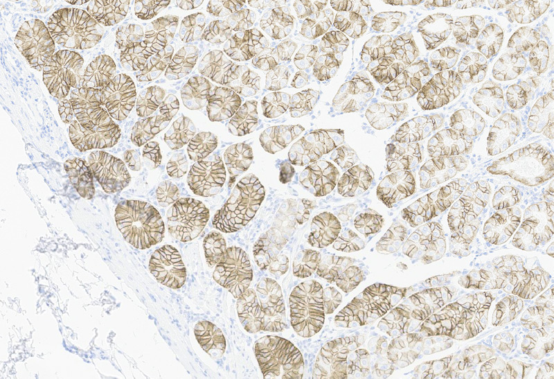
Dako link 48-10X

Dako link 48-40X

Dako link 48-40X
> Using Claudin-18.2 primary antibody (Cat. No. HCS-S278) to stain normal gastric tissues, positive cell cytoplasm and/or cell membrane specific positive staining (staining intensity ≥ 1), no non-specific staining (staining intensity < 1) , no background staining (staining intensity < 1 ).
Performance comparison
Titer: ACRO at 1/1000 dilution and Competitor A at 1/200 dilution, color intensity is similar

Claudin18.2 (ACRO)
1:1000-40X

Claudin18.2 (ACRO)1:1000-40X

Claudin18.2 (Competitor A)
1:200-40X
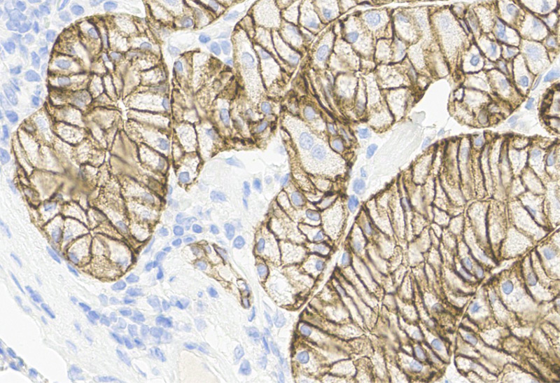
Claudin18.2 (Competitor A)1:200-40X

Claudin18.2 (Competitor A)
1:500-40X
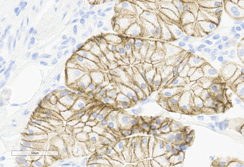
Claudin18.2 (Competitor A)1:500-40X
> Immunohistochemical analysis of paraffin-embedded human stomach tissue labeling Claudin-18.2 with ACRO/Competitor A. Membranous staining on human gastric epithelial cells is observed. Counter stained with Hematoxylin. Perform heat mediated antigen retrieval with Tris/EDTA buffer pH 9.0 before commencing with IHC staining protocol.
Sensitivity: ACRO staining intensity is stronger than Competitor A

Claudin18.2 (ACRO)
1:1000-4X

Claudin18.2 (ACRO)1:1000-4X

Claudin18.2 (Competitor A)
1:200-4X
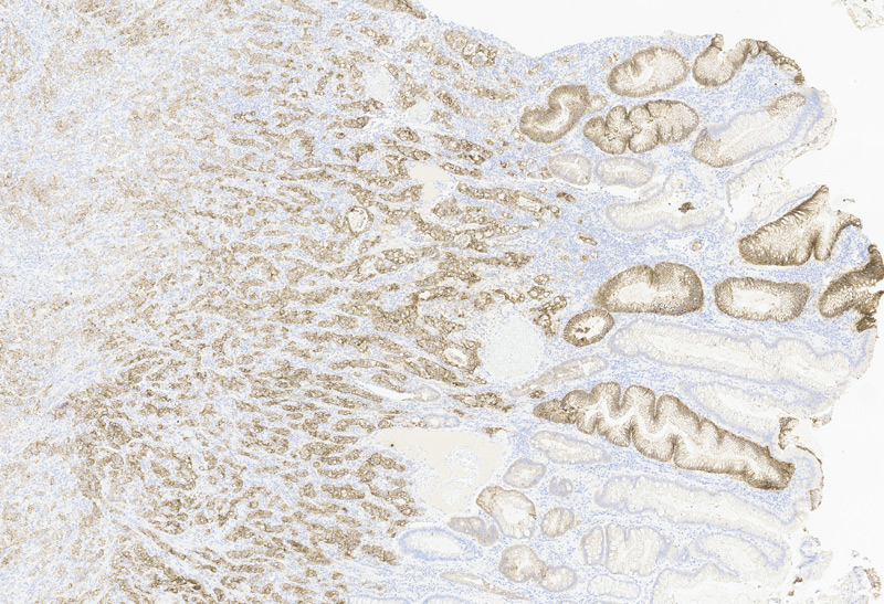
Claudin18.2 (Competitor A)1:200-4X

Claudin18.2 (Competitor A)
1:500-4X
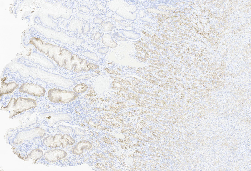
Claudin18.2 (Competitor A)1:500-4X

Claudin18.2 (ACRO)
1:1000-40X
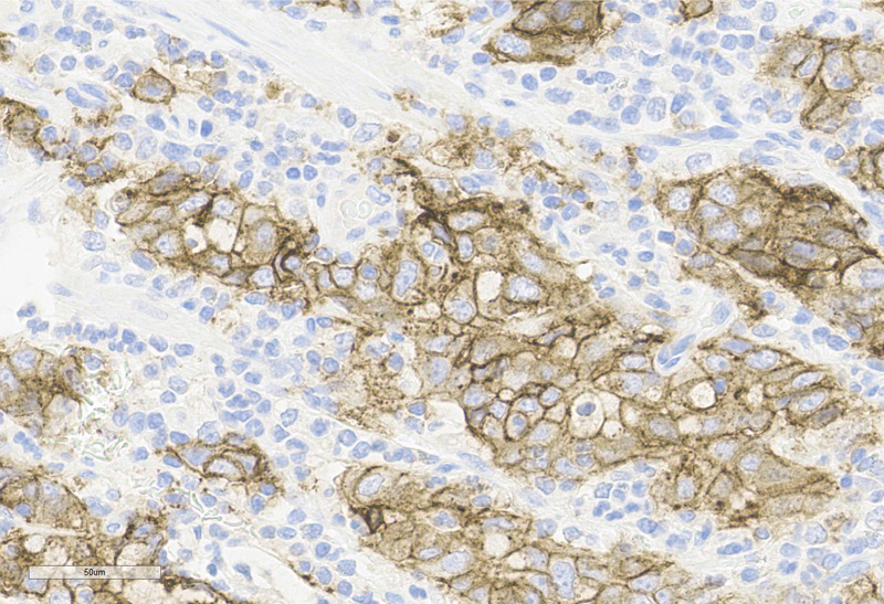
Claudin18.2 (ACRO)1:1000-40X

Claudin18.2 (Competitor A)
1:200-40X
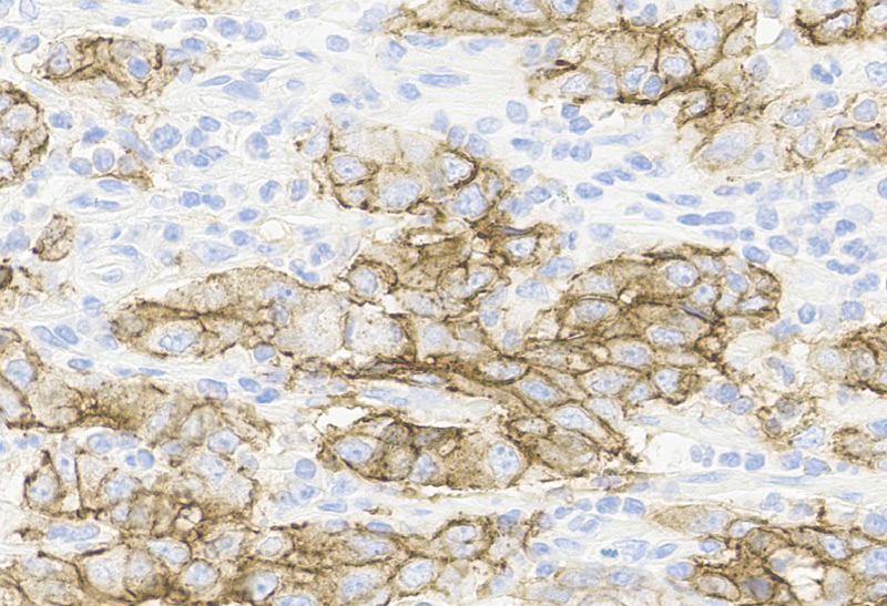
Claudin18.2 (Competitor A)1:200-40X

Claudin18.2 (Competitor A)
1:500-40X
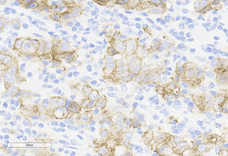
Claudin18.2 (Competitor A)1:500-40X
> Immunohistochemical analysis of paraffin-embedded human gastric cancer labeling Claudin-18.2 with ACRO/Competitor A. Membranous staining on tumor cells is observed. Counter stained with Hematoxylin. Perform heat mediated antigen retrieval with Tris/EDTA buffer pH 9.0 before commencing with IHC staining protocol.
ACRO Quality

This web search service is supported by Google Inc.

totop

Laisser un message